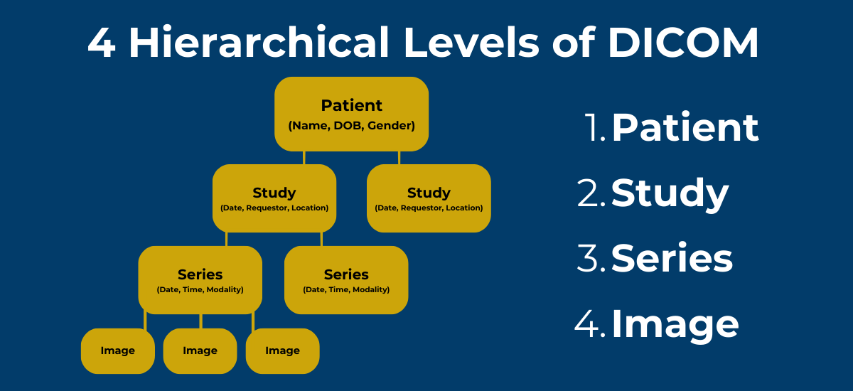
The 4 Hierarchical Levels of DICOM

Digital Imaging and Communications in Medicine (DICOM) is the global standard image processing model used by health care organizations and medical professionals. Prevalent in radiology, radiotherapy and cardiology, DICOM systems simplify image management. They organize information into four levels — patient, study, series and image.
What Is DICOM and Why Does It Matter?
DICOM is the standard file format for recording, storing and transmitting visual medical records. Created in 1993, it is now the primary image format for hospitals, clinical practices and other medical facilities. Various platforms, like a Picture Archiving and Communications System (PACS), use DICOM files to structure, save and transfer patient information.
As an industry standard, DICOM enables you to manage medical images seamlessly. You can share relevant medical records without converting to other file formats. You can also handle diverse data forms, including neurophysiology waveforms, ultrasound and radiation results, sonar records, virtual reality and augmented reality datasets. DICOM supports and integrates with different modalities, or health care devices, to retrieve, archive, send and process high-quality files.
What Are the 4 Hierarchies of DICOM?

Knowing how this industry-standard file formatting assembles data through the DICOM hierarchy is the key to using it effectively. In software, a hierarchy is the diagrammatic model used to organize information. Hierarchies structure information into main folders and subcategories. DICOM’s hierarchy consists of four categories — patient, study, series and image. Every file processed using DICOM organizes information into these four levels, creating a data pattern that clarifies medical administration and clinical file management.
The four hierarchies of DICOM include:
- Patient
- Study
- Series
- Image
Patient
The first level of your DICOM file records the patient’s information. It stores patient case details and demographics, including their name, date of birth, age and gender. For every image processed via DICOM, you can read the corresponding patient information.
Study
A study refers to the patient exam. DICOM organizes studies as imaging procedures a patient undergoes. Patients can have multiple procedures, so they can have one or more studies. Data recorded under the study tier include:
- Date: The date of the scheduled procedure.
- Time: Time when the examiner performed the procedure.
- Requester: The medical practitioner who requested the exam.
- Modality: Type of procedure or device.
- Location: The practice or hospital where the patient underwent the exam.
Series
One study can include several series. These are variations of the exam, scans or images.
During a procedure, patients may be scanned several times using the same machine. Multiple physical scans can make up a series in one study. The second series type involves digital reconstruction. You may reconstruct one CT scan in assorted ways, rendering different images. These variations form a series within that singular CT study. Details recorded for each series include:
- Date: The date of the exam.
- Time: The image's retrieval time or when the exam occurred.
- Modality: The type of scan or device used.
Image
The different images in a series are the fourth level of the hierarchy of DICOM. Every series can include one or more images, which are also called instances. Patient, study and series information adds context to these medical images or instances.
Find the Right Image Management Tools With Candelis, Inc.
Interpreting DICOM files is essential for providing advanced patient care. As the industry-preferred mode for medical image processing, working with DICOM offers several benefits. You can comply with industry and legal standards on information processing, optimize your facility workflows and data administration, and improve diagnostic accuracy.
Candelis, Inc. offers ImageViewTM DICOM viewers with state-of-the-art medical software for evaluating CR/DR records, ultrasounds, MRI images and more. We’ve designed each solution with unique and intuitive features to fit your needs. Use them to improve diagnostics, enhance care accuracy, speed up image viewing and optimize patient outcomes.
Book a free demo with us for an image-viewing solution that elevates your practice.
- Log in to post comments
