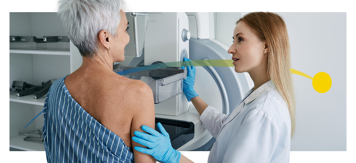
The History of Mammography and Mammograms

More than 42 million mammograms were reported in the United States in 2024, with regular screenings helping slash the risk of dying from breast cancer almost in half. But how did we get here? While the mammogram was technically invented in the early 20th century, breast cancer screening went through several evolutions to become what it is today.
Who Invented the Mammogram?
In the early 19th century, breast study was only conducted through X-ray units intended for other diagnostic procedures. This resulted in excessive radiation exposure. Image quality was also poor due to the lack of compression and ability to focus on specific spots.
That's not to say that breast disease wasn't a topic of study — German surgeon Albert Salomon published work discussing the use of radiology on breast disease to differentiate between benign and malignant entities in 1913. He is considered the inventor of the mammogram machine and used X-ray technology to demonstrate how different types of breast cancer spread through lymph nodes.
The Early Work: The 1930s to 1950s
Stafford Warren performed the first mammograph in the U.S. in 1930. Warren's work analyzed the potential role of mammograms as a preoperative measure for assessing tumors and established a baseline for the regular appearance of the mammary gland. He also emphasized the need to examine both breasts for better comparison to detect and identify abnormalities.
The next major development in the mammography history timeline happened in 1949. Raul Leborgne, a radiologist from Uruguay, worked to enhance image quality and showcased how patient positioning and different radiological parameters made a difference.
Leborgne also worked on differential diagnosis between benign and malignant using microcalcifications. He developed the breast compression technique to lower radiation exposure and improve image quality, which is still used today.
Pathologist Helen Ingleby developed a study in 1950 that assessed breast characteristics and how they related to a woman's age, menstrual status and other factors. She also developed a cross-sectional histological technique for diagnosis.
Screening Becomes Standard: The 1960s and 1970s
Mammograms started to become standard practice in the 1960s and 70s. In 1962, Robert Egan — considered a pioneer in mammography — reported the first-time detection of more than 50 cases of occult breast cancer after conducting 2,000 mammograms.
Around the same time, researchers demonstrated the role of mammographic studies in private clinics, helping standardize the practice. The American College of Radiology (ACR) created centers across the country for training, which eventually developed into the ACR Mammography Committee.
Charles Gross developed the first unit mammography-specific apparatus in 1965. It provided better contrast between various parts of the breast and had a built-in compression system.
Mammograph Units See Improvement
Breast cancer was a leading cause of mortality in the U.S. by the 1970s. The medical science community continued exploring safer and more effective screening options, like the use of screens and films, to help reduce overall radiation exposure.
In 1974, several publications called attention to mammography's role in diagnosing some forms of cancer. Just two years later, improvements were made to the system for marking nonpalpable breast lesions, now using a needle and wire system that would eventually become the Kopans wires still used today.
Work was soon published regarding mammography magnification and the need to evolve existing apparatuses to reflect recent findings in the field. Breast tumor diagnosis as a whole was becoming more holistic and comprehensive, now integrating more indirect and subtle signs.
By 1978, the moving grid was introduced, which scattered radiation and improved image contrast. This led to better mammography equipment and more detailed images. Around this time, mobile mammography apparatuses were also gaining traction.
The Rise of Digital: The 1980s to Modern Day

By the mid-1980s, László Tabár and collaborators reported a significant decrease in mortality rates after analyzing screenings of more than 130,000 women. In 1994, U.S. President Bill Clinton proclaimed the third Friday in October as National Mammography Day, falling during Breast Cancer Awareness Month.
There was also a new emphasis on digital mammography to build on this momentum. Just two years later, the Food and Drug Administration (FDA) published guidelines on how to get approval for commercial digital mammography equipment, bringing it into the mainstream.
The FDA approved in 2000 the Senographe 2000D, the first full-field digital mammography (FFDM) system that worked similarly to traditional apparatuses but used direct digital acquisition of the mammography images.
In 2011, the FDA went on to approve digital breast tomosynthesis (DBT) — an advanced form of mammography that captures images from multiple angles and creates a more detailed image of the breast.
Breast Cancer Screening Today
Screening for breast cancer is more effective than ever — there is a 40% reduction in mortality in women 40 to 84 who screen yearly versus those who don't. Recent efforts to make mammographs more accessible by integrating them directly into OB-GYN practices have seen great success and could be instrumental in bridging the gap between those who schedule annual screenings and those who can't due to location or time.
Tools like artificial intelligence (AI) for results analyses and liquid biopsy for predictive genomic information are still in their early stages. However, they are also promising indicators of the future of breast cancer screenings.
The Evolution of Screening Recommendations
As mammography technology improved and we've expanded our understanding of breast cancer diagnosis, official breast cancer screening recommendations have also changed. Before the 1980s, screening was recommended for women as young as 35 if she, her mother or her sister had a history of breast cancer. Women 50 and over were recommended to have yearly mammograms.
By 1997, guidelines became much more generalized — all women 40 and over were recommended for annual screening. This standard persisted through the 2000s and early 2010s. In October 2015, guidelines evolved into what health care providers still use today:
- Women 40 to 44 should have the choice to begin annual screenings, given they've considered the potential risks.
- Women 45 to 54 should have annual screenings.
- Women 55 and older have the choice to continue screening yearly. They may also transition to biyearly mammograms as long as they are in good health and expected to live at least 10 more years.
From History to Innovation: Transform Mammography for Every Patient
Though mammographs have become a highly effective, lifesaving tool, there's still work to be done to improve diagnosis accuracy and enhance the patient experience. Practices face challenges like lengthy download times for images, ultimately reducing the number of studies radiologists can review daily and delaying critical diagnoses.
The Candelis Advanced Breast Imaging PACS addresses these issues through enhanced image quality and tools. These can help you provide a more thorough diagnosis with a backend server designed to optimize storage and image movement. This improves overall efficiency and frees radiologists to analyze more studies per hour so you can provide faster, more accurate results.
Book a free customized demo today and help pioneer the next era in mammogram history.

- Log in to post comments
